Image of the Week — April 15, 2018
CIL:39063 — http://www.cellimagelibrary.org/images/39063
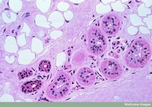
Description: Light micrograph of a ‘semithin’ section (1.5 µm thick) of the dermis layer of skin from a monkey. The upper left side shows an area of (empty) fat cells surrounded by collagen fibers. The lower right side contains six sections of a coiled sweat gland and three excretory ducts. The columnar cells forming the wall of the sweat gland secrete the solutes, these are interdispersed with myosecretory cells (triangular red shapes particularly visible in gland at far right) that can contract to squeeze the secretion out of the lumen of the gland into the excretory duct. The section was stained with haematoxylin and eosin (H+E), which is a common histological stain added to tissue sections to distinguish cells.
Author: Spike Walker
Licensing: Attribution-NonCommercial-NoDerivs 2.0 UK: England & Wales (CC BY-NC-ND 2.0 UK)
Archive link


Комментариев нет:
Отправить комментарий