What a Relief
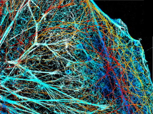
Nothing in life is completely flat. Even our thinnest cells are speckled with contours and bumps – tiny 3D structures that might be crucial to their health, but difficult to spot. Here, a combination of super resolution microscopy and mathematics reveals the structure of a mammalian cell’s cytoskeleton – the mesh of protein ‘bones’ that keep cell membranes propped up. Each is artificially coloured by its vertical height – with lighter shades of blue and yellow being furthest from the cell’s floor – over a distance two million times smaller than the average tent. The techniques use information about how patterns of light are usually ‘seen’ by the microscope to remove some of the blurriness common when picturing such small structures – revealing more about how cells move and adhere among tissues in the body, but also how they might be targeted with medical treatments.
Written by John Ankers
- Image from work by Clément Cabriel and colleagues
- Institut des Sciences Moléculaires d’Orsay, CNRS, Univ. Paris-Sud, Université Paris-Saclay, Orsay Cedex, France
- Image originally published under a Creative Commons Licence (BY 4.0)
- Published in Nature Communications, April 2019
You can also follow BPoD on Instagram, Twitter and Facebook
Archive link


Комментариев нет:
Отправить комментарий