Image of the Week – June 17, 2019
CIL:199 — http://cellimagelibrary.org/images/199
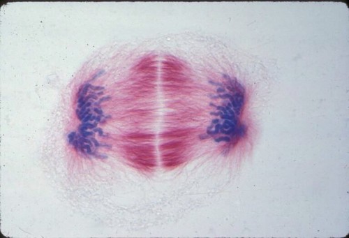
Description: Lily mitosis. A light microscope image of a cell from the endosperm of an African globe lily Haemanthus (Scadoxus) katherinae. This is one frame of a sequence that shows all phases of mitosis. The lily is considered a good organism for studying cell division because its endosperm has a liquid phase and chromosomes are thick and easier to see than human ones. Staining shows microtubules in red and chromosomes in blue. This image showing a cell in telophase is the 5th of a grouped series that spans mitosis.
Author: Andrew S. Bajer
Licensing: Public Domain: This image is in the public domain and thus free of any copyright restrictions. However, as is the norm in scientific publishing and as a matter of courtesy, any user should credit the content provider for any public or private use of this image whenever possible.
The Spectacular World of Brain Imaging Flipbook — http://go.technologynetworks.com/the-spectacular-world-of-brain-imaging-flipbook
Archive link


Комментариев нет:
Отправить комментарий