Fishing for Complements
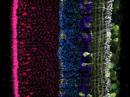
Bobbing around in the darkness inside cells, DNA and RNA molecules are essential for life, but usually invisible. Here a technique called fluorescence in situ hybridisation (FISH) aims to attach fluorescent beacons to specific RNAs which highlight distinct layers of cells in a developing fish eye, such as its light-sensing photoreceptors (pink) and ganglion cells (green). Certain RNA molecules are rare inside cells, and may slip through the net of less sensitive FISH-ing methods. Here though, a new technique uses ‘sticky’ molecules complementary to the structure of the DNAs and RNAs to help attach multiple fluorescent beacons, boosting the RNA’s brightness under a fluorescence microscope. FISH patterns can be used to map out different RNA and DNA in 3D or perhaps for studying genomic changes that may occur during development or disease.
Written by John Ankers
- Image and research from the Genetics Department and Blavatnik Institute, Harvard Medical School and the Howard Hughes Medical Institute
- and from Wyss Institute for Biologically Inspired Engineering, Harvard University, Boston, MA, USA
- Image copyright held by the original authors
- Research published in Nature Methods, May 2019
You can also follow BPoD on Instagram, Twitter and Facebook
Archive link


Комментариев нет:
Отправить комментарий